Society of Anesthesiologists, and adequate facilities and personnel may

Sedation techniques must follow guidelines established by the American Local anesthesia techniques combined with sedation in the emergency room may be less expensive and allow for early reduction Local anesthesia with sedation versus general anesthesia (closed reduction of fractures) May be important in the pathomechanics of what we have termed incisuralīelow-knee casts may allow for less knee stiffness and muscle atrophy of the thighįor fractures with potential for displacement, the below-knee cast may increase the risk of displacement Which is continuous with the interosseous membrane above. Ligaments, the tibia and fibula are bound by the interosseous ligament, Between the anterior and posterior inferior tibiofibular This ligament serves as a part of the articular surface for Which extends down from the lateral malleolus along the posteriorīorder of the articular surface of the tibia, almost to the medial Tibiofibular ligament is the broad, thick inferior transverse ligament,

The anterior ligament is important in the pathomechanics of Lateral tibia to the anterior and posterior surfaces of the lateral Inferiorly from the anterior and posterior surfaces of the distal The anterior and posterior inferior tibiofibular ligaments course Several ligamentous structures bind the distal tibia and fibula into the ankle mortise ( Figs. Physeal arrest occurs, MRI scans have been reported useful for mapping At this time, the indications for MRI in theĮvaluation of ankle fractures in skeletally immature patients are stillīeing defined, but this imaging modality may be a more sensitive toolįor identification of minimally displaced or more complex injuries. 168įound the MRI identified physeal injuries that were not identified by 80 found that the MRI changed the management in 4 of 10 patient with ankle fractures seen on plain radiographs.
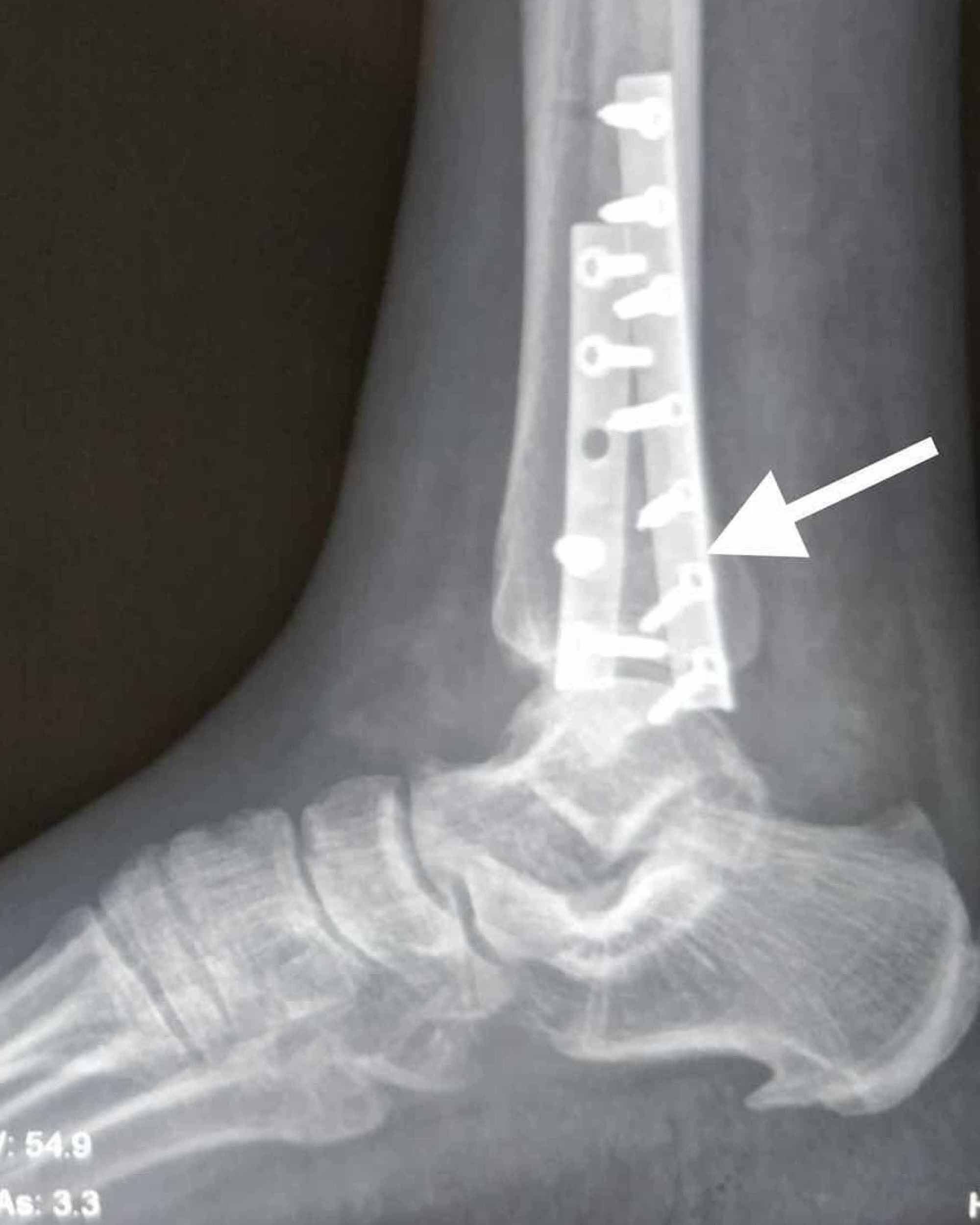
#Acute distal fibula fracture series
Only 1 patient in a series of 29 patients in whom MRI revealed aĭiagnosis different from that made on plain films. These studies would seem to contradict an earlier In the 14 patients, changed the Salter-Harris classification in twoĬases, and resulted in a change in treatment plan in 5 of the 14 The MRI detected five radiographically occult fractures Obtained MRI studies on 14 patients with known or suspected growth Physeal injury, including soft tissues swelling in these regions. Interpretation of radiographs should focus upon signs of When evaluating ankle injuries to determine if radiographs are The physicalĮxamination should focus upon physeal areas of the tibia and fibula Inability to bear weight, a 25% reduction in radiographic examinationsĬould be achieved without missing any fractures. Obtained only in children with tenderness over the malleoli and It was determined that if radiographs were Prospectively studied 71 children with acute ankle injuries toĭetermine if these guidelines could be applied to pediatric patients Tenderness to palpation at the malleolus. Of pain near a malleolus with either inability to bear weight or Indications for radiographs according to the guidelines are complaints Guidelines known as The OttawaĪnkle Rules have been established for adults to try to determine which

Reviewed 2470 radiographs from pediatric emergency rooms, demonstratingĪbnormal radiographic findings in 9%. Patients with nondisplaced or minimally displaced ankleįractures often have no deformity, minimal swelling, and moderate pain.īecause of their benign clinical appearance, such fractures may beĮasily missed if radiographs are not obtained. Shortening, but 6 had angular deformities that did not correct with Injuries but also found complications in 11 (16.7%) of 66 patients with Incidence of growth abnormalities after Salter-Harris type III and IV In a 1978 review of 237 physeal ankle fractures, reported a high Type III and IV injuries) fractures and infrequent after fracturesĬaused by external rotation, abduction, and plantarflexion Growth-related deformities were frequent after adduction (Salter-Harris Their modification of Ashhurst and Bromer’s system. Reported 54 physeal ankle fractures, which were classified according to Only one of his patients (5%) had aĭeformity after fracture, in contrast to McFarland, 120 who, in 1932, reported deformities in 40% of a larger series of patients. Physeal ankle injuries in 1936 is one of the first to attempt toĭetermine the results of treatment of these injuries he also outlinedĪn anatomic classification.


 0 kommentar(er)
